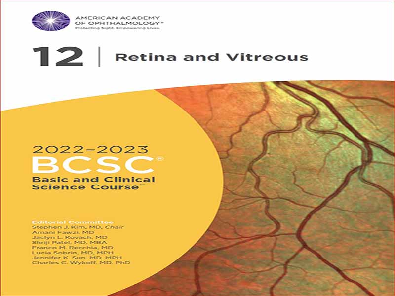
- عنوان کتاب: Retina and Vitreous 2022–2023 BCSC
- نویسنده: Stephen J. Kim, MD
- حوزه: چشم پزشکی
- سال انتشار: 2022
- تعداد صفحه: 529
- زبان اصلی: انگلیسی
- نوع فایل: pdf
- حجم فایل: 18.4 مگابایت
BCSC بخش 12، Ret ina و Vitreous، به 3 قسمت مرتب شده است. بخش اول، مبانی و رویکردهای تشخیصی، آناتومی، تصویربرداری و ارزیابی عملکردی را در 3 فصل پوشش میدهد. فصل 1 یک نمای کلی از آناتومی بخش خلفی ارائه می دهد. فصل 2 تکنیک های معاینه را مورد بحث قرار می دهد، از جمله بیومیکروسکوپی با لامپ شکاف متسع در ترکیب با لنزهای غیر تماسی یا تماسی پیش قرنیه، و همچنین افتالموسکوپی غیرمستقیم، که اغلب با فرورفتگی اسکلرال برای تسهیل مشاهده رت اینا قدامی کمک می کند. مستندسازی یافتههای بالینی، چه از طریق توصیف و چه با تصویر، یک عنصر ضروری در معاینه کامل بخش خلفی است. آزمایشهای جانبی مانند عکاسی فوندوس، اتوفلورسانس، آنژیوگرافی فلورسین، آنژیوگرافی سبز ایندوسیانین، توموگرافی انسجام نوری (OCT)، افتالموسکوپی لیزری اسکن، میکروپریمتری و الکتروفیزیولوژی ممکن است اطلاعات تشخیصی بیشتری ارائه دهد یا به تشخیص تغییرات در طول زمان کمک کند. تجسم های مختلف OCT (حوزه زمانی، دامنه طیفی و منبع جابجایی) شایسته ذکر ویژه است. OCT شاید به متداول ترین آزمایش کمکی چشم پزشکی تبدیل شده است و درک ما را از بسیاری از شرایط ویترئورت متحول می کند. سونوگرافی (که اکووگرافی نیز نامیده می شود) با استفاده از هر دو تکنیک اسکن A و B، برای معاینه بیماران با محیط غیر شفاف مفید است، می تواند در تشخیص ضایعات شبکیه یا مشیمیه کمک کند و می تواند برای نظارت بر اندازه ضایعه استفاده شود. فصل 3 به بررسی تست های الکتروفیزیولوژیک و اهمیت آنها در تشخیص و پیگیری بیماری شبکیه می پردازد. بخش دوم، بیماریهای شبکیه و زجاجیه، که شامل فصلهای 4 تا 17 است، بیماریهای خاص و آسیبهای وارده به بخش خلفی را پوشش میدهد. تکنیک های تشخیصی مناسب در سراسر این بحث ها نشان داده شده است. بر حسب ضرورت، یافتههای مصور مرتبط با بیماریهای شیشهای توصیفشده بهعنوان نکات برجسته – با استفاده از طرحوارهها و همچنین فناوریهای تصویربرداری مختلف- به جای مجموعهای جامع ارائه میشوند. روایتهایی که مدیریت و درمان بیماریهای شبکیه تحت پوشش بخش دوم را توصیف میکنند، با توصیف کارآزماییهای بالینی مهمی تکمیل میشوند که به ایجاد شیوههای مبتنی بر شواهد مناسب کمک کردهاند. به عنوان بخشی از بازنگری عمده کتاب، محتوای قسمت دوم مجدداً ایجاد شده است. بیماری مادرزادی و ثابت شبکیه، که در فصلی جداگانه در آخرین ویرایش مورد بحث قرار گرفت، اکنون بخشی از فصل 12، “بیماری شبکیه ارثی و کوروئید” است. همچنین، فصلی با عنوان «بیماریهای رابط زجاجیه و ویترئورت» که قبلاً بعد از «جدایی مجدد و ضایعات مستعدکننده» ظاهر میشد، اکنون مقدم بر آن است و جریان منطقیتری ارائه میدهد.
BCSC Section 12, Ret ina and Vitreous, is arranged into 3 parts. Part I, Fundamentals and Diagnostic Approaches, covers ret i nal anatomy, imaging, and functional evaluation in 3 chapters. Chapter 1 provides an overview of the anatomy of the posterior segment. Chapter 2 discusses examination techniques, including dilated slit- lamp biomicroscopy in combination with precorneal non– contact or contact lenses, as well as indirect ophthalmoscopy, which is often aided by scleral indentation to facilitate viewing the anterior ret ina. Documenting clinical findings, either by description or by illustration, remains an essential ele ment of a complete posterior segment examination. Ancillary testing, such as fundus photography, autofluorescence, fluorescein angiography, indocyanine green angiography, optical coherence tomography (OCT), scanning laser ophthalmoscopy, microperimetry, and electrophysiology, may provide additional diagnostic information or help detect changes over time. The vari ous incarnations of OCT (time- domain, spectraldomain, and swept- source) deserve special mention. OCT has become perhaps the most commonly ordered ancillary ophthalmic test and is revolutionizing our understanding of many vitreoret i nal conditions. Ultrasonography (also called echography) employing both A- and B- scan techniques, is useful for examining patients with opaque media, can aid in the diagnosis of ret i nal or choroidal lesions, and can be used to monitor a lesion’s size. Chapter 3 reviews electrophysiologic tests and their significance in diagnosis and followup of ret i nal disease. Part II, Diseases of the Ret ina and Vitreous, which includes Chapters 4 through 17, covers specific diseases of and trauma to the posterior segment. Appropriate diagnostic techniques are indicated throughout these discussions. By necessity, the illustrative findings associated with the described vitreoret i nal diseases are presented as highlights— using schematics as well as the dif fer ent imaging technologies— rather than as a comprehensive collection. Narratives describing the management of, and therapy for, the ret i nal diseases covered in Part II are complemented by descriptions of the impor tant clinical trials that have helped establish appropriate evidence- based practices. As part of the major revision of the book, content in Part II has been reor ga nized. Congenital and stationary ret i nal disease, discussed in a separate chapter in the last edition, is now part of Chapter 12, “Hereditary Ret i nal and Choroidal Disease.” Also, the chapter titled “Diseases of the Vitreous and Vitreoret i nal Interface,” which previously appeared after “Ret i nal Detachment and Predisposing Lesions,” now precedes it, providing a more logical flow.
این کتاب را میتوانید از لینک زیر بصورت رایگان دانلود کنید:
Download: Retina and Vitreous 2022–2023 BCSC


نظرات کاربران