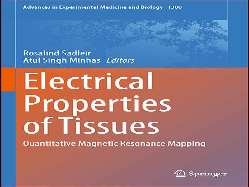
- عنوان کتاب: Electrical Properties of Tissues
- نویسنده: Rosalind Sadleir
- حوزه: مهندسی پزشکی
- سال انتشار: 2022
- تعداد صفحه: 211
- زبان اصلی: انگلیسی
- نوع فایل: pdf
- حجم فایل: 7.55 مگابایت
خواص الکتریکی بافت ها یک ویژگی اساسی است که بینشی منحصر به فرد و بسیار حساس را در مورد ترکیب و عملکرد آنها ارائه می دهد. در حالی که مقادیر رسانایی و گذردهی ذاتی بافت ممکن است با استفاده از نمونههای بریده شده یا در محل با استفاده از سلولهای رسانایی یا کاوشگر تعیین شود، علاقه روزافزونی به تصویربرداری از توزیع این ویژگیها در بدن وجود دارد. حوزه توموگرافی امپدانس الکتریکی (EIT) اکنون بیش از 40 سال است که وجود دارد و کاربردهای مهمی در زمینه هایی مانند نظارت غیرتهاجمی ریه و تصویربرداری عملکردی دارد. اگرچه EIT یک تکنیک نظارتی بسیار حساس است، اما محدودیتها به دلیل نامطلوب بودن مشکل معکوس به وجود میآیند. تصویربرداری چگالی فعلی با استفاده از تصویربرداری رزونانس مغناطیسی برای اولین بار توسط اسکات و جوی که در دانشگاه تورنتو در اوایل دهه 1990 کار می کردند، نشان داده شد. پیشنهاد استفاده از این رویکرد برای توزیع رسانایی تصویر نیز توسط این گروه ارائه شد. اولین رویکردها برای تصویربرداری از چگالی جریان و رسانایی با فرض سه اندازهگیری متفاوت، نشان دهنده سه جزء توزیع چگالی شار مغناطیسی ناشی از جریان خارجی است. در اوایل دهه 2000، سئو و وو به رویکردی کمک کردند که شامل اندازهگیری و استفاده از تنها یک جزء منفرد از چگالی شار مغناطیسی بود. تقریباً در همان زمان، Katscher و همکارانش در حال توسعه استراتژیهای عملی برای تصویربرداری توزیع رسانایی و گذردهی در فرکانسهای Larmor سیستمهای MRI بودند. در طول سال ها، روش های متعددی برای بازسازی میدان های الکترومغناطیسی و توزیع های رسانایی پیشنهاد و آزمایش شده است. جریان های مورد نیاز برای تصویربرداری از رسانایی در پایین ترین فرکانس ها از بیش از 20 میلی آمپر به حدود 1.5 میلی آمپر کاهش یافته است و اندازه گیری رسانایی سر انسان در داخل بدن به دست آمده است. اندازهگیریهای هدایت فرکانس بالا در آزمایشهای بالینی در حال آزمایش هستند، و جدیدترین روشهای بازسازی رسانایی به هیچ وجه شامل هیچ تجویز فعلی نمیشوند و فقط شامل تصویربرداری تانسور انتشار و توزیعهای هدایت فرکانس بالا میشوند. این منطقه همچنین از علاقه به مطالعه مکانیسمها و درمانهای فردی در درمانهای تعدیل عصبی مانند تحریک AC و DC ترانس جمجمهای بهره میبرد که نیاز به اندازهگیری چگالی جریان و توزیع میدان الکتریکی دارد. این جلد با فرض اینکه خواننده یک دانشجوی کارشناسی یا کارشناسی ارشد با تمرکز بر موضوعات پیشرفته در MRI باشد، یا محققی است که با حوزه کلی خواص الکتریکی بافت و MRI آشنا نیست، سازماندهی شده است. فصل 1 با یک درمان اساسی از عوامل زیربنایی خواص الکتریکی بافت ها آغاز می شود. مقدمهای عملی برای تحلیل اجزای محدود و مدلسازی الکترومغناطیسی، که معمولاً در طول بازسازی مورد نیاز است، در فصل آورده شده است. 2. فصل 3 مقدمه ای اساسی برای MRI را پوشش می دهد که بر سخت افزار، فیزیک، عبارات ریاضی برای فضای k، توالی پالس، بازسازی تصویر و روش های SNR تمرکز می کند، که اساس تصویربرداری فاز در MRI را تشکیل می دهد. فصلهای 4 و 5 به ساخت فانتومها برای تصویربرداری رسانایی مربوط میشوند و دو فصل بعدی یک مرور کلی از روشهای MRI برای بازسازی چگالی جریان (فصل 6) و هدایت (فصل 7) ارائه میکنند. این جلد با بررسی عوامل مهم در اندازه گیری رسانایی فرکانس بالا به پایان می رسد (فصل 8). این کتاب حاصل کارگاه موفقی است که در سی و نهمین کنفرانس بینالمللی سالانه کنفرانس مهندسی پزشکی و زیستشناسی IEEE که در جزیره ججو، جمهوری کره در سال 2018 برگزار شد. مایلیم از دوستان و دوستان خود تشکر و قدردانی کنیم. مربیان Eung Je Woo، Jin Keun Seo، و Oh In Kwon. نویسندگان همکار ما کاملیا گابریل، مونیش چاوهان، ساوراو ز. ک. ساجیب، اولریش کاتچر، روت الیور، و نیتیش کاتچ؛ و خانواده ها و همکاران ما برای مشارکت در این پروژه. ما همچنین میخواهیم از مری استابر و دیپاک راوی در اسپرینگر برای کمک آنها در گردآوری این جلد تشکر کنیم.
The electrical properties of tissues are a fundamental characteristic providing a unique and extremely sensitive insight into their composition and function. While values for intrinsic tissue conductivity and permittivity may be determined using excised samples or in situ using conductivity cells or probes, there is increasing interest in imaging the distribution of these properties within the body. The area of electrical impedance tomography (EIT) has now existed for over 40 years and has important applications in areas such as non-invasive lung monitoring and functional imaging. Though EIT is a very sensitive monitoring technique, limitations arise because of the general illposedness of the inverse problem. Current density imaging using magnetic resonance imaging was first demonstrated by Scott and Joy, working at the University of Toronto in the early 1990s. The suggestion to use this approach to image conductivity distributions was also made by this group. The earliest approaches to imaging both current density and conductivity proceeded assuming three different measurements, representing the three components of the magnetic flux density distribution caused by an external current. In the early 2000s, Seo and Woo contributed to an approach that involved measuring and using only a single component of the magnetic flux density. At around the same time, Katscher and colleagues were developing practical strategies for imaging conductivity and permittivity distributions at the Larmor frequencies of MRI systems. Over the years, numerous methods for reconstructing electromagnetic fields and conductivity distributions have been suggested and tested. The currents required to image conductivities at the lowest frequencies have reduced from over 20mA to around 1.5mA, and measurements of human head conductivities have been obtained in vivo. High-frequency conductivity measurements are being tested in clinical trials, and the newest conductivity reconstructionmethods involve no current administration at all, involving only diffusion tensor imaging and high-frequency conductivity distributions. The area is also benefiting from interest in studying mechanisms and individualized treatments in neuromodulation therapies such as transcranial AC and DC stimulation, which require measurement of current density and electric field distributions. The volume is organized assuming the reader is an undergraduate or graduate student focusing on advanced topics in MRI, or a researcher unfamiliar with the general area of tissue electrical properties and MRI. Chapter 1 begins with a basic treatment of the factors underlying the electrical properties of tissues. A practical introduction to the finite element analysis and electromagnetic modeling, typically required during reconstructions, is given in Chap. 2. Chapter 3 covers a basic introduction to MRI focusing on hardware, physics, mathematical expressions for k-space, pulse sequences, image reconstruction, and SNR methods, forming the basis of phase imaging in MRI. Chapters 4 and 5 are concerned with constructing phantoms for conductivity imaging, and the next two chapters present a comprehensive overview ofMRI methods for reconstructing current density (Chap. 6) and conductivity (Chap. 7). The volume concludes with an examination of important factors in measuring high-frequency conductivity (Chap. 8). This book arose out of a successful workshop that was held at the 39th Annual International Conference of the IEEE Engineering in Medicine and Biology conference which took place at Jeju Island, Republic of Korea, in 2018.We would like to thank and acknowledge our friends and mentors Eung Je Woo, Jin Keun Seo, and Oh In Kwon; our co-authors Camelia Gabriel, Munish Chauhan, Saurav Z. K. Sajib,UlrichKatscher, Ruth Oliver, and Nitish Katoch; and our families and colleagues for their contributions to this project. We would also like to convey our gratitude to Merry Stuber and Deepak Ravi at Springer for their assistance in compiling this volume.
این کتاب را میتوانید از لینک زیر بصورت رایگان دانلود کنید:
Download: Electrical Properties of Tissues


نظرات کاربران