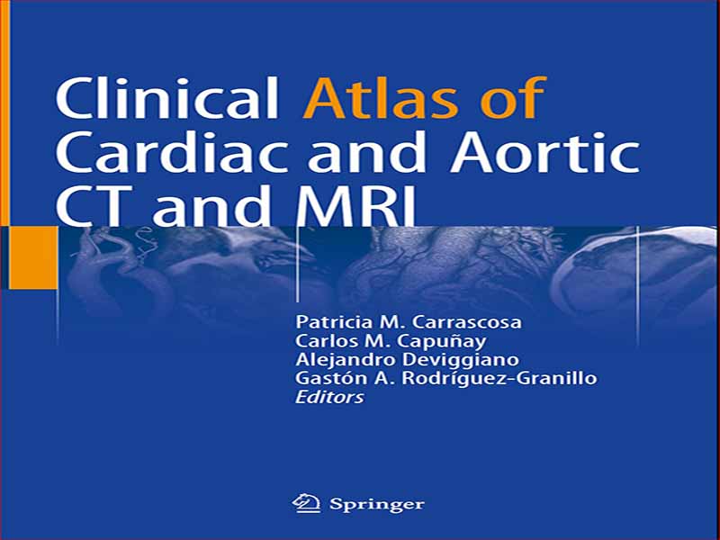
- عنوان کتاب: Clinical Atlas of Cardiac and Aortic CT and MRI
- نویسنده: Patricia M. Carrascosa
- حوزه: قلب و عروق, MRI
- سال انتشار: 2019
- تعداد صفحه: 373
- زبان اصلی: انگلیسی
- نوع فایل: pdf
- حجم فایل: 45.7 مگابایت
در چند دهه گذشته، CT و MRI قلب به سرعت از ابزارهای تحقیقاتی جذاب، به استراتژیهای تشخیصی کمکی، و در نهایت به نقش اساسی در طیف وسیعی از بیماریهای ایسکمیک و غیرسکمیک تغییر کردهاند. به موازات آن، بیماری ساختاری قلب، که ارتباط نزدیکی با افزایش اخیر چندین مداخله از راه پوست غیرکرونری دارد، هم از نظر تعداد روشها و هم از نظر تنوع مداخلات، رشد زیادی داشته است. در واقع، این تکنیکهای تصویربرداری انقلابی در رویکرد تشخیصی و درمانی بیماران قلبی عروقی ایجاد میکنند و به تدریج به تخصصهای فرعی جذاب در میان رادیولوژیستها و متخصصان قلب تبدیل میشوند. در عمل معمول بالینی، CT و/یا MRI قلب و آئورت برای تعیین تشخیص در میان بیماران با یافتههای غیرقابل قطعی، آزمایشهای غیرتشخیصی، بیماریهای نادر، برای بهبود ویژگیهای موجودات خاص مانند کاردیومیوپاتیها و/یا به عنوان راهنمایی و نظارت بر مداخلات پوستی انجام میشود. مانند تعویض دریچه آئورت ترانس کاتتر و ترمیم آئورت اندوواسکولار و غیره. در نتیجه، متخصصان قلب و/یا رادیولوژیستهایی که در ارزیابی این بیماران دخیل هستند، مرتباً با موارد غیرعادی چالش برانگیز مواجه میشوند. علاوه بر این، چنین پزشکانی معمولاً در هر دو زمینه تصویربرداری دخالت دارند. بر اساس دلایل ذکر شده در بالا، به جای کتب درسی گسترده و با جزئیات فنی، نیازی برآورده نشده برای متنی مختصر، خاص و مبتنی بر گزارش موردی وجود دارد که هدف آن توصیف یافتههای اصلی، ویژگیهای کلیدی و متداولترین گونههایی است که میتوان در این مقاله یافت. به صورت روزانه برای این منظور، نمونهای کامل از شایعترین و غیرمعمولترین مواردی را که در تمرین معمول یک مرکز تصویربرداری (Diagnostico Maipu، بوئنوس آیرس، آرژانتین) ارزیابی میشوند، جمعآوری کردیم، که در زمینه بالینی آنها ارائه شده است. علاوه بر این، کارشناسان مشهور از سایر موسسات موارد استثنایی اضافی، به ویژه بیماریهای نادر مانند بیماری قلبی مادرزادی و تومورهای قلبی را ارائه میدهند. موارد در 9 فصل اصلی زیر مرتب شده اند: (1) آناتومی قلب و ناهنجاری های کرونر. (2) کاردیومیوپاتی ایسکمیک. (3) انفارکتوس میوکارد با عروق کرونر غیر انسدادی. (4) کاردیومیوپاتی غیرسکمیک. (5) بیماری ساختاری قلب و راهنمایی رویههای از راه پوست. (6) بیماری قلبی مادرزادی. (7) توده های قلبی و تومورها. (8) بیماری پریکارد. و (9) بیماری آئورت.
In the past few decades, cardiac CT and MRI have rapidly shifted from appealing research tools, to adjuvant diagnostic strategies, and ultimately to a fundamental role in a wide range of ischemic and nonischemic diseases. In parallel, structural heart disease, closely related to the recent surge of several noncoronary percutaneous interventions, has underwent an enormous growth both in terms of number of procedures and variety of interventions. Indeed, these imaging techniques are revolutionizing the diagnostic and therapeutic approach of cardiovascular patients and progressively becoming attractive subspecialties among radiologists and cardiologists. In routine clinical practice, cardiac and aortic CT and/or MRI are performed to define diagnosis among patients with inconclusive findings, nondiagnostic tests, rare diseases, to improve characterization of specific entities such as cardiomyopathies, and/or as guidance and surveillance of percutaneous interventions such as transcatheter aortic valve replacement and endovascular aortic repair, among others. Consequently, cardiologists and/or radiologists involved in the evaluation of these patients are regularly confronted to challenging unusual cases. Furthermore, such physicians are usually implicated in both imaging fields. Based on the abovementioned reasons, rather than extensive and technically detailed textbooks, there is an unmet need for a concise, specific, case-report-based text aimed at describing the main findings, key features, and most frequent variants that can be found on a daily basis. For this purpose, we collected a thorough sample of the most common and uncommon cases that are evaluated in routine practice of an imaging facility (Diagnostico Maipu, Buenos Aires, Argentina), provided in their clinical context. Moreover, renowned experts from other institutions provide additional exceptional cases, particularly involving rare diseases such as congenital heart disease and cardiac tumors. Cases are arranged within the following nine main chapters: (1) Cardiac Anatomy and Coronary Anomalies; (2) Ischemic Cardiomyopathy; (3) Myocardial Infarction with Nonobstructive Coronary Arteries; (4) Nonischemic Cardiomyopathy; (5) Structural Heart Disease and Guidance of Percutaneous Procedures; (6) Congenital Heart Disease; (7) Cardiac Masses and Tumors; (8) Pericardial Disease; and (9) Aortic Disease.
این کتاب را میتوانید بصورت رایگان از لینک زیر دانلود نمایید.








نظرات کاربران