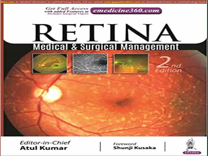
- عنوان کتاب: Retina – Medical and Surgical Management
- نویسنده: Atul Kumar
- حوزه: بیماری چشم
- سال انتشار: 2022
- تعداد صفحه: 724
- زبان اصلی: انگلیسی
- نوع فایل: pdf
- حجم فایل: 43.3 مگابایت
برای من بسیار خوشحالم که دومین نسخه به روز شده شبکیه چشم: مدیریت پزشکی و جراحی را ارائه می کنم. این به روز رسانی های اخیر و آخرین پیشرفت ها در تشخیص، دستورالعمل های مدیریت بالینی، ابزار دقیق و جراحی های شبکیه چشم را در بر می گیرد و دیدگاهی جامع در مورد فوق تخصص شبکیه در اختیار خوانندگان قرار می دهد که برای خوانندگان بسیار مفید خواهد بود. این امر پس از ماهها گردآوری دادهها، جمعآوری تصویر و بازبینی متن با ورودیهای گسترده از دستیاران ارشد و ساکنان خردسال که در مرکز RP، AIIMS، دهلی نو، هند کار میکنند، امکانپذیر شده است. در سال 2016 بود که ایده یک کتاب درسی جامع در مورد شبکیه چشم برای دستیاران و متخصصین شبکیه چشم و همچنین یک کتاب مرجع برای ورق زدن روی میز چشم پزشکان شاغل به ذهنم رسید و بنابراین تصمیم گرفتم افکار و ایده های خود را در مورد همه بیاورم. آنچه را که از 30 سال گذشته تاکنون هر روز می بینم و انجام می دهم در قالب یک کتاب. من از روزهای اقامتم در مرکز RP همیشه علاقه زیادی به شبکیه چشم به عنوان یک فوق تخصص داشتم و در مورد این رشته از بیماری های شبکیه چشم شگفت زده بودم، جایی که تغییرات سریع در فناوری با تکنیک های جدیدتر مطابقت دارد و به سرعت تکامل می یابد و خود را به یک عمل پیچیده پزشکی چشم ادامه می دهد. و جراحی که از طیف وسیعی از روش های تشخیصی و درمانی برای درمان بیماری های چشمی استفاده می کند. از گزارشهای موردی گرفته تا فاز 3 کارآزماییهای بالینی چند مرکزی، دادههای منتشر شده همچنان به صورت تصاعدی انباشته میشوند. در نتیجه برای پزشکانی که با بیماری شبکیهای شبکیهای سر و کار دارند، چه عمومی و چه متخصص، بهطور فزایندهای دشوار است که اطلاعات جمعآوریشده را برای مراقبت از بیمار به کار ببرند. این کتاب درسی در درجه اول در نظر گرفته شده است تا دستیاران چشم پزشکی و چشم پزشکان جامع را با منبعی به روز و مبتنی بر بالینی ارائه دهد که طیف کاملی از بیماری های زجاجیه شبکیه پزشکی و جراحی را پوشش می دهد. متخصصان فوق تخصص در این زمینه نیز باید آن را به عنوان یک مرجع بررسی و انتخاب مفید بدانند. اگرچه، این متن به معنای جامع بودن است، ما سعی کردهایم جنبههای اساسی و مهم بالینی عمل پزشکی و جراحی شبکیهای را برجسته کنیم. فصل مربوط به وضعیت بیماری بر ویژگی های بالینی، تشخیص و مدیریت تاکید دارد. کل فصل ها به رایج ترین مشکلاتی که با آن مواجه می شوند اختصاص داده شده است. بخشهای جدیدی در مورد نقش نوظهور جراحی شبکیه شبکیه سه بعدی «heads up» و توموگرافی انسجام نوری یکپارچه با میکروسکوپ در جراحیهای شبکیه اضافه شده است. متن به 9 بخش تقسیم شده است که هر بخش دارای فصول زیادی است که به شیوه ای سیستماتیک به بیماری های مختلف شبکیه و علائم، علائم و مدیریت آنها اختصاص دارد. جدیدترین پلتفرم های تصویربرداری با تصاویر و تکنیک های جراحی در این کتاب درسی شرح داده شده است. گردآوری یک کتاب درسی که سعی میکند فصلها را هم از نظر محتوایی جاری و هم با جزئیات همراه با طرحبندی یکسان داشته باشد، تلاش ویژه همه مشارکتکنندگان را برای رعایت دستورالعملهای دقیق میطلبد و از همه کسانی که برای مشارکت در نگارش این کتاب درسی دعوت شدهاند تشکر میکنم. تلاش های قابل تحسین درباره کتاب: مقدمه این کتاب به عنوان یک کتاب درسی و راهنمای عمیق برای درک موضوع شبکیه و زجاجیه و بیماریهای مرتبط با آن با تصاویر رنگی خوب و تمام بهروزرسانیهای اخیر در نظر گرفته شده است. به طور کلی به 9 بخش با فصل های مرتبط در هر بخش تقسیم شده است. بخش 1 به علوم پایه و تشخیص اختصاص دارد و آناتومی و فیزیولوژی مفید چشم، تصویربرداری مهم شبکیه با جدیدترین پلتفرمهای تصویربرداری و تصاویر واضح از بیماریهای شبکیه را که با آخرین سختافزار با فناوری پیشرفته تصویربرداری شدهاند را پوشش میدهد. الکتروفیزیولوژی چشم و USG چشمی که اطلاعات مربوط به اختلالات رتینوکوروئیدی را ارائه می دهد این بخش را کامل می کند. بخش 2 به دژنراسیون شبکیه و دیستروفی های فوندال اختصاص داده شده است. این شامل نزدیک بینی پاتولوژیک و ضایعات آن با نمایش تصویری، رتینیت پیگمانتوزا و سندرم های مرتبط، ویترو رتینوپاتی و دیستروفی ارثی شبکیه و مشیمیه است. بخش 3 به طور کامل به بیماری های ماکولا از جمله کوریورتینوپاتی سروزی مرکزی (CSC)، دژنراسیون ماکولا وابسته به سن (AMD) و علل منتهی به غشای نئوواسکولار مشیمیه ثانویه (CNVM) اختصاص دارد. بیماریهای واسط ویترئومولی و کشش ویترئوماکولار (VMT) در ادامه توضیح داده میشوند، و هر کدام به طور دقیق فصلی را به غشاهای اپی رتینال (ERMs) و سوراخهای ماکولا اختصاص میدهند. ماکولوپاتی کششی نزدیکبین اکنون بیشتر در چشمهای نزدیکبین بهویژه با دستگاه توموگرافی منسجم نوری (SS-OCT) تصویربرداری میشود و در ادامه توضیح داده میشود و پس از آن ماکولوپاتی حفرهای دیسک بینایی و مدیریت آن با پر کردن غشای محدود کننده داخلی (ILM) توضیح داده میشود.
It gives me great pleasure to bring out the second updated edition of the Retina: Medical and Surgical Management. It encompasses the recent updates and latest advances in diagnostics, clinical management guidelines, instrumentation and vitreoretinal surgeries and will provide the readers a holistic view about the retinal subspecialty which will prove to be extremely useful to the readers. This has been possible after months of data compilation, image acquisition and revision of text with extensive inputs from senior residents and junior residents working at RP Centre, AIIMS, New Delhi, India. It was in 2016 that I conceived the idea of a comprehensive textbook on retina for residents and vitreoretinal fellows, and also a reference book to flip through, lying on the desk of the practicing ophthalmologists and so decided to put down my thoughts and ideas of all what I see and do daily since the last 30 years into a book form. I have always been passionate about retina as a sub-specialty since my residency days at RP Centre and was amazed about this field of vitreoretinal diseases where rapid changes in technology meets newer techniques and continues to evolve rapidly expanding itself into a complex practice of ophthalmic medicine and surgery that utilizes a wide range of diagnostic and therapeutic modalities to treat ocular diseases. From case reports to phase 3 multicenter clinical trials, published data continues to accumulate in exponential fashion. As a result it is increasingly difficult for the clinician dealing with vitreoretinal disease, both general and specialist alike to apply the amassed information to patient care. This textbook is primarily intended to provide the ophthalmology residents and practicing comprehensive ophthalmologists with an up-to-date, clinically oriented source that covers the full spectrum of medical and surgical vitreoretinal disease. Subspecialists in the field should also find it useful as a review and selected reference. Although, this text is meant to be comprehensive, we have tried to highlight the essential, clinically important aspects of vitreoretinal medical and surgical practice. Chapter dealing with disease state emphasize clinical feature, diagnosis, and management. Entire chapters are devoted to the most commonly encountered problems. New sections about emerging role of 3D ‘heads up’ vitreoretinal surgery and microscope integrated optical coherence tomography in retinal surgeries have been added. The text is divided into 9 sections, each section having many chapters in a systematic fashion devoted to the various retinal diseases and their signs, symptoms and management. The latest imaging platforms with illustrations and surgical techniques have been described in this textbook. Assembling a textbook that attempts to have the chapters both current and detailed in content along with uniform layout requires a special effort from all the contributors to adhere to strict guidelines and I am grateful to all those invited to participate in the writing of this textbook for their applaudable efforts. About the Book: An Introduction This book is intended to serve as an in-depth textbook and guide for understanding the subject of retina and vitreous and the diseases related to it with well-illustrated colored images and all the recent updates. It is broadly divided into 9 sections with relevant chapters in each section. Section 1 is dedicated to basic sciences and diagnostics and covers useful anatomy and physiology of the eye, the all important retinal imaging with the latest imaging platforms and crisp pictures of retinal diseases imaged with the latest high technology hardware. Ocular electrophysiology and ophthalmic USG which provides information of retinochoroidal disorders completes this section. Section 2 is devoted to retinal degenerations and fundal dystrophies. This includes pathological myopia and its lesions with pictorial representation, retinitis pigmentosa and related syndromes, hereditary retinal and choroidal vitreoretinopathy and dystrophies. Section 3 is totally devoted to macular diseases including central serous chorioretinopathy (CSC), age-related macular degeneration (AMD), and etiologies leading to secondary choroidal neovascular membrane (CNVM). Vitreomacular interface diseases and vitreomacular traction (VMT) are explained next, closely followed by a chapter each devoted to epiretinal membranes (ERMs), and macular holes each. Myopic traction maculopathy is now imaged more commonly in myopic eyes especially with the swept-source optical coherence tomography (SS-OCT) device and is next described, followed by optic disc pit maculopathy and its management with internal limiting membrane (ILM) stuffing.
این کتاب را میتوانید بصورت رایگان از لینک زیر دانلود نمایید.
Download: Retina – Medical and Surgical Management


نظرات کاربران