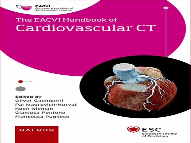
- عنوان کتاب: The European Society of Cardiology Series Oliver
- نویسنده: Oliver Gaemperli
- حوزه: قلب و عروق
- سال انتشار: 2023
- تعداد صفحه: 386
- زبان اصلی: انگلیسی
- نوع فایل: pdf
- حجم فایل: 62.4 مگابایت
از زمان معرفی توموگرافی کامپیوتری (CT) در سال 969 توسط سر گادفری هانسفیلد، این فناوری با سرعتی خیره کننده تکامل یافته و به سنگ بنای تصویربرداری غیرتهاجمی در عمل بالینی تبدیل شده است. جهشی عظیم با معرفی اسکنرهای سی تی اسکن چند تکه با زمان چرخش کوتاه و وضوح مکانی و زمانی لازم برای حل کوچکترین قسمتهای متحرک آناتومی قلب انجام شد: شریانهای کرونر. با اصلاحات بیشتر تکنولوژیکی، شواهد کارآزمایی بالینی و توصیههای راهنما، CT قلب، و مهمتر از همه، CT آنژیوگرافی عروق کرونر (CCTA)، به عنوان یک روش تصویربرداری غیرتهاجمی قلب ضروری و یک آزمایش خط اول مهم برای بیماری عروق کرونر مورد استقبال قرار گرفته است. اخیراً، پتانسیل CT قلبی در تشخیص و راهنمایی درمان در انواع دیگر آسیبشناسیهای قلبی فراتر از بیماری عروق کرونر، از جمله بیماری دریچهای، فیبریلاسیون دهلیزی و سایر آریتمیها، اندوکاردیت، تودههای قلبی، کاردیومیوپاتیها و غیره مشهود شده است. بر این اساس، انجمن اروپایی تصویربرداری قلب و عروق (EACVI) اعلام کرده است که یکی از مهمترین اولویت های آنها تسهیل آموزش و آموزش در زمینه CT قلب و عروق از طریق دوره های آموزشی، کنگره ها و یک برنامه صدور گواهینامه ساختاریافته است (به فصل 4. مراجعه کنید). این کتاب راهنما گامی مهم در جهت انتشار مهارت ها و دانش در سی تی قلب و عروق است. این به عنوان یک همراه مختصر و عملی در نظر گرفته شده است که برای دانش آموزان، کارآموزان یا کاربران پیشرفته (متخصصان قلب، رادیولوژیست ها، جراحان قلب و تکنسین ها) در تمرین روزمره خود مفید است. چهار بخش گسترده شامل جنبه های فنی و زمینه فیزیکی، اندیکاسیون های عروق کرونر (مانند CCTA، تصویربرداری تصلب شرایین، استنت ها و بای پس ها، و تصویربرداری سی تی عملکردی)، اندیکاسیون های غیر کرونر (CT برای بیماری دریچه، اندوکاردیت عفونی، CT دهلیز چپ، مادرزادی است. بیماری های قلبی، توده های قلبی، یافته های خارج از قلب و غیره) و در نهایت آموزش و شایستگی در CT قلب. این کتاب دارای فصلهای کوتاه است که با تصاویر، جداول و خلاصههای فشرده فراوان غنی شده است که دسترسی سریع و بصری را تسهیل میکند. ما معتقدیم که در میان بسیاری از کتابهای درسی موجود، کتاب راهنمای ما یک شکاف مهم را پر میکند، و امیدواریم که راه خود را در کتهای آزمایشگاهی بسیاری از پزشکان پیدا کند. از زمان معرفی توموگرافی کامپیوتری (CT) در سال 969 توسط سر گادفری هانسفیلد، این فناوری با سرعتی خیره کننده تکامل یافته و به سنگ بنای تصویربرداری غیرتهاجمی در عمل بالینی تبدیل شده است. جهشی عظیم با معرفی اسکنرهای سی تی اسکن چند تکه با زمان چرخش کوتاه و وضوح مکانی و زمانی لازم برای حل کوچکترین قسمتهای متحرک آناتومی قلب انجام شد: شریانهای کرونر. با اصلاحات بیشتر تکنولوژیکی، شواهد کارآزمایی بالینی و توصیههای راهنما، CT قلب، و مهمتر از همه، CT آنژیوگرافی عروق کرونر (CCTA)، به عنوان یک روش تصویربرداری غیرتهاجمی قلب ضروری و یک آزمایش خط اول مهم برای بیماری عروق کرونر مورد استقبال قرار گرفته است. اخیراً، پتانسیل CT قلبی در تشخیص و راهنمایی درمان در انواع دیگر آسیبشناسیهای قلبی فراتر از بیماری عروق کرونر، از جمله بیماری دریچهای، فیبریلاسیون دهلیزی و سایر آریتمیها، اندوکاردیت، تودههای قلبی، کاردیومیوپاتیها و غیره مشهود شده است. بر این اساس، انجمن اروپایی تصویربرداری قلب و عروق (EACVI) اعلام کرده است که یکی از مهمترین اولویت های آنها تسهیل آموزش و آموزش در زمینه CT قلب و عروق از طریق دوره های آموزشی، کنگره ها و یک برنامه صدور گواهینامه ساختاریافته است (به فصل 4. مراجعه کنید). این کتاب راهنما گامی مهم در جهت انتشار مهارت ها و دانش در سی تی قلب و عروق است. این به عنوان یک همراه مختصر و عملی در نظر گرفته شده است که برای دانش آموزان، کارآموزان یا کاربران پیشرفته (متخصصان قلب، رادیولوژیست ها، جراحان قلب و تکنسین ها) در تمرین روزمره خود مفید است. چهار بخش گسترده شامل جنبه های فنی و زمینه فیزیکی، اندیکاسیون های عروق کرونر (مانند CCTA، تصویربرداری تصلب شرایین، استنت ها و بای پس ها، و تصویربرداری سی تی عملکردی)، اندیکاسیون های غیر کرونر (CT برای بیماری دریچه، اندوکاردیت عفونی، CT دهلیز چپ، مادرزادی است. بیماری های قلبی، توده های قلبی، یافته های خارج از قلب و غیره) و در نهایت آموزش و شایستگی در CT قلب. کتاب راهنما دارای فصلهای کوتاه است که با تصاویر، جداول و خلاصههای فشرده فراوان غنی شده است که دسترسی سریع و شهودی را تسهیل میکند. ما معتقدیم که در میان بسیاری از کتابهای درسی موجود، کتاب راهنمای ما یک شکاف مهم را پر میکند، و امیدواریم که راه خود را در کتهای آزمایشگاهی بسیاری از پزشکان پیدا کند.
Since the introduction of computed tomography (CT) in 969 by Sir Godfrey Hounsfield, this technology has evolved at a breathtaking pace to become a cornerstone of non- invasive imaging in clinical practice. A giant leap was realized with the introduction of multislice CT scanners with short rotation times and the necessary spatial and temporal resolution to resolve the smallest, moving parts of cardiac anatomy: the coronary arteries. Supported by further technological refinements, clinical trial evidence, and guideline recommendations, cardiac CT, and, foremost, coronary CT angiography (CCTA), has been embraced as an indispensable noninvasive cardiac imaging modality and an important first- line test for coronary artery disease. Recently, the potential of cardiac CT has become evident in the diagnosis and guidance of treatment in a variety of other cardiac pathologies beyond coronary artery disease, including valvular disease, atrial fibrillation and other arrhythmias, endocarditis, cardiac masses, cardiomyopathies, and others. On these grounds, the European Association of Cardiovascular Imaging (EACVI) has declared that one of their foremost priorities is to facilitate education and training in cardiovascular CT through teaching courses, congresses, and a structured certification programme (see Chapter 4.). This handbook represents an important step towards the dissemination of skills and knowledge in cardiovascular CT. It is conceived as a concise and practical companion, to benefit students, trainees, or advanced users (cardiologists, radiologists, cardiac surgeons, and technicians) in their everyday practice. Four broad sections cover the technical aspects and physical background, coronary indications (e.g. CCTA, atherosclerosis imaging, stents and bypasses, and functional CT imaging), non- coronary indications (CT for valve disease, infective endocarditis, CT of the left atrium, congenital heart disease, cardiac masses, extracardiac findings, etc.), and, finally, training and competence in cardiac CT. The handbook features short chapters, enriched with plenty of illustrations, tables, and condensed summaries, which facilitate rapid and intuitive access. We believe that among the many textbooks available, our handbook fills an important gap, and hope that it will find its way into the pockets of many practitioners’ lab coats. Since the introduction of computed tomography (CT) in 969 by Sir Godfrey Hounsfield, this technology has evolved at a breathtaking pace to become a cornerstone of non- invasive imaging in clinical practice. A giant leap was realized with the introduction of multislice CT scanners with short rotation times and the necessary spatial and temporal resolution to resolve the smallest, moving parts of cardiac anatomy: the coronary arteries. Supported by further technological refinements, clinical trial evidence, and guideline recommendations, cardiac CT, and, foremost, coronary CT angiography (CCTA), has been embraced as an indispensable noninvasive cardiac imaging modality and an important first- line test for coronary artery disease. Recently, the potential of cardiac CT has become evident in the diagnosis and guidance of treatment in a variety of other cardiac pathologies beyond coronary artery disease, including valvular disease, atrial fibrillation and other arrhythmias, endocarditis, cardiac masses, cardiomyopathies, and others. On these grounds, the European Association of Cardiovascular Imaging (EACVI) has declared that one of their foremost priorities is to facilitate education and training in cardiovascular CT through teaching courses, congresses, and a structured certification programme (see Chapter 4.). This handbook represents an important step towards the dissemination of skills and knowledge in cardiovascular CT. It is conceived as a concise and practical companion, to benefit students, trainees, or advanced users (cardiologists, radiologists, cardiac surgeons, and technicians) in their everyday practice. Four broad sections cover the technical aspects and physical background, coronary indications (e.g. CCTA, atherosclerosis imaging, stents and bypasses, and functional CT imaging), non- coronary indications (CT for valve disease, infective endocarditis, CT of the left atrium, congenital heart disease, cardiac masses, extracardiac findings, etc.), and, finally, training and competence in cardiac CT. The handbook features short chapters, enriched with plenty of illustrations, tables, and condensed summaries, which facilitate rapid and intuitive access. We believe that among the many textbooks available, our handbook fills an important gap, and hope that it will find its way into the pockets of many practitioners’ lab coats.
این کتاب را میتوانید بصورت رایگان از لینک زیر دانلود نمایید.


نظرات کاربران