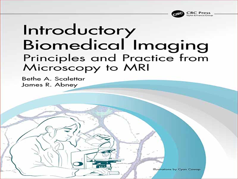
- عنوان کتاب: Introductory Biomedical Imaging
- نویسنده: Bethe-A.-Scalettar
- حوزه: زیست پزشکی
- سال انتشار: 2023
- تعداد صفحه: 338
- زبان اصلی: انگلیسی
- نوع فایل: pdf
- حجم فایل: 38.7 مگابایت
تصویربرداری همه جا هست. ما از چشمانمان برای دیدن و از دوربین ها برای عکس گرفتن استفاده می کنیم. دانشمندان از میکروسکوپ و تلسکوپ برای نگاه کردن به سلول ها و خارج شدن به فضا استفاده می کنند. پزشکان از اولتراسوند، اشعه ایکس، رادیوایزوتوپ ها و ام آر آی برای بررسی داخل بدن ما استفاده می کنند. اگر در مورد تصویربرداری کنجکاو هستید، این کتاب درسی را باز کنید تا اصول اولیه را بیاموزید.
تصویربرداری ابزاری قدرتمند در تحقیقات علمی بنیادی و کاربردی است و همچنین نقش مهمی در تشخیص، درمان و تحقیقات پزشکی دارد. این کتاب درسی در مقطع کارشناسی، تکنیک های تصویربرداری پیشرفته و فیزیک زیربنایی آنها را معرفی می کند. مفاهیم اولیه از الکترومغناطیس، اپتیک و فیزیک مدرن برای توضیح اشکال برجسته میکروسکوپ نوری، و همچنین آندوسکوپی، اولتراسوند، رادیوگرافی پروجکشن و توموگرافی کامپیوتری، تصویربرداری رادیونوکلئید، و تصویربرداری تشدید مغناطیسی استفاده میشوند. این کتاب درسی همچنین پردازش و تجزیه و تحلیل تصویر دیجیتال را پوشش می دهد. اصول نظری با مشکلات مشروح، برنامهها، فعالیتها و آزمایشها و با تأکید بر مضامین تکرار شونده، از جمله تأثیر وضوح، کنتراست و نویز بر کیفیت تصویر، تقویت میشوند. خوانندگان اصول تصویربرداری، قابلیت های تشخیصی و نقاط قوت و ضعف تکنیک ها را یاد خواهند گرفت.
این کتاب درسی پیدایش خود را داشت، و در دوره “تصویربرداری زیست پزشکی” در کالج لوئیس و کلارک در پورتلند، OR بررسی شده است، و برای تسهیل تدریس دوره های مشابه در سایر موسسات طراحی شده است. این در پوشش میکروسکوپ نوری و تصویربرداری پزشکی در سطح متوسط منحصر به فرد است و در پوشش مواد در چندین سطح از پیچیدگی استثنایی است.
Imaging is everywhere. We use our eyes to see and cameras to take pictures. Scientists use microscopes and telescopes to peer into cells and out to space. Doctors use ultrasound, X-rays, radioisotopes, and MRI to look inside our bodies. If you are curious about imaging, open this textbook to learn the fundamentals.
Imaging is a powerful tool in fundamental and applied scientific research and also plays a crucial role in medical diagnostics, treatment, and research. This undergraduate textbook introduces cutting-edge imaging techniques and the physics underlying them. Elementary concepts from electromagnetism, optics, and modern physics are used to explain prominent forms of light microscopy, as well as endoscopy, ultrasound, projection radiography and computed tomography, radionuclide imaging, and magnetic resonance imaging. This textbook also covers digital image processing and analysis. Theoretical principles are reinforced with illustrative homework problems, applications, activities, and experiments, and by emphasizing recurring themes, including the effects of resolution, contrast, and noise on image quality. Readers will learn imaging fundamentals, diagnostic capabilities, and strengths and weaknesses of techniques.
This textbook had its genesis, and has been vetted, in a “Biomedical Imaging” course at Lewis & Clark College in Portland, OR, and is designed to facilitate the teaching of similar courses at other institutions. It is unique in its coverage of both optical microscopy and medical imaging at an intermediate level, and exceptional in its coverage of material at several levels of sophistication.
این کتاب را میتوانید از لینک زیر بصورت رایگان دانلود کنید:
Download: Introductory Biomedical Imaging






نظرات کاربران