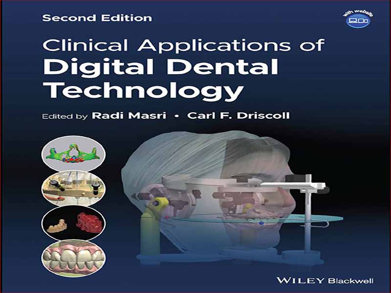
- عنوان کتاب: Clinical Applications of Digital Dental Technology
- نویسنده: Radi Masri, BDS, MS, PhD
- حوزه: دندانپزشکی
- سال انتشار: 2023
- تعداد صفحه: 313
- زبان اصلی: انگلیسی
- نوع فایل: pdf
- حجم فایل: 20.6 مگابایت
از زمانی که اولین تصاویر رادیوگرافی داخل دهانی توسط دندانپزشک آلمانی، Otto Walkhoff (Langland و همکاران 1984) در اوایل سال 1896، تنها 14 روز پس از W.C، در معرض دید قرار گرفت، تصویربرداری به یک شکل در حرفه دندانپزشکی مورد استفاده قرار گرفت. رونتگن علناً کشف اشعه ایکس خود را اعلام کرد (McCoy 1919، Bushong 2008). در طول تاریخ بیش از 120 ساله رادیوگرافی دهان، پیشرفتهای مهم بسیاری انجام شده است (انجمن رادیولوژیستهای دهان و فک و صورت آمریکا (AAOMR) 2021، مولتنی 2021). اولین گیرنده ها شیشه بودند، اما فیلم استاندارد را برای بخش عمده قرن بیستم تا دهه 1990 تعیین کرد، زمانی که توسعه رادیوگرافی دیجیتال برای استفاده دندانپزشکی توسط شرکت Trophy تجاری شد که سیستم RVGui را منتشر کرد (Mouyen et al. 1989). . شرکت های دیگری مانند کداک، جندکس، شیک، پلانمکا، سیرونا و دکسیس نیز پیشگامان اولیه رادیوگرافی دیجیتال بودند. پذیرش رادیوگرافی دیجیتال توسط حرفه دندانپزشکی آهسته اما پیوسته بوده است و به نظر می رسد که حداقل تا حدی توسط نظریه “اشاعه نوآوری” که توسط دکتر اورت راجرز (2003) حمایت شده است، کنترل شده است. کار او توضیح میدهد که چگونه پیشرفتهای تکنولوژیکی مختلف توسط کاربران نهایی فناوری در نیمه دوم قرن بیستم و اوایل قرن بیست و یکم اتخاذ شده است. دو مورد از مهمترین اصول پذیرش فناوری، مفاهیم آستانه و جرم بحرانی است. آستانه یک ویژگی یک گروه است و به تعداد افراد یک گروه اشاره دارد که باید از یک فناوری استفاده کنند یا قبل از اینکه یک فرد فناوری را بپذیرد یا در یک فعالیت شرکت کند، باید در یک فعالیت شرکت کنند. توده بحرانی یکی دیگر از ویژگی های یک گروه است و در نقطه ای از زمان رخ می دهد که افراد کافی در گروه یک نوآوری را پذیرفته اند تا امکان رشد خودپایه آینده پذیرش نوآوری را فراهم کنند. همانطور که نوآوران بیشتری از یک فناوری مانند رادیوگرافی دیجیتال استفاده می کنند، مزیت درک شده از فناوری بیشتر و بیشتر می شود تا تعداد روزافزونی از سایر پذیرندگان آینده تا زمانی که این فناوری در نهایت رایج شود. رادیوگرافی دیجیتال رایج ترین فناوری پیشرفته دندانپزشکی است که بیماران در طول ویزیت های تشخیصی تجربه می کنند. طبق آخرین نظرسنجی مطب دندانپزشکی که توسط کنفرانس مدیران برنامه کنترل پرتو در سالهای 2014-2015 انجام شد و در سال 2019 منتشر شد، 86 درصد مطبها از تصویربرداری دیجیتال استفاده میکردند (کنفرانس مدیران برنامه کنترل تشعشع 2019). تعداد دندانپزشکانی که از تصویربرداری دیجیتال استفاده می کنند همچنان در حال افزایش است. اگر هنوز از فیلم استفاده میکنید، این سوال نباید این باشد که “آیا باید به سیستم رادیوگرافی دیجیتال روی بیاورم؟” اما در عوض “کدام سیستم دیجیتال به راحتی در دفتر من ادغام می شود؟” این منجر به سوال دیگری می شود – رادیوگرافی دیجیتال در مقایسه با ادامه استفاده از فیلم معمولی چه مزایایی برای حرفه دندانپزشکی ارائه می دهد؟ اجازه دهید به آنها نگاه کنیم.
رایجترین کلاس سرعت یا حساسیت فیلم داخل دهانی فیلم سرعت D است. نمونه بارز این فیلم در ایالات متحده، شورای ملی با سرعت فوق العاده کداک در حفاظت و اندازه گیری تشعشع (NCRP 2012) است. مقدار دوز تشعشع مورد نیاز برای تولید یک تصویر تشخیصی با استفاده از این فیلم تقریباً دو برابر مقدار مورد نیاز برای Kodak’s Insight، یک فیلم با سرعت F است. به عبارت دیگر سرعت فیلم F دو برابر سرعت فیلم D است. به گفته مویال، که از دادههای NEXT در سال 1999 استفاده کرد، دوز ورودی پوست برای گاز گرفتن خلفی با سرعت D ~1.7 میلیگری است (Moyal 2007). طبق گزارش NCRP #172، دوز متوسط ورودی پوست برای قرار گرفتن در معرض فیلم با سرعت D ~ 2.2 میلی گی است، در حالی که دوز معمول فیلم با سرعت E-F ~ 1.3 میلی گی است و دوز متوسط ورودی پوست از سیستم های دیجیتال ~ است. 0.8 میلی گی (NCRP 2012). با توجه به NCRP #145 و دیگران، به نظر می رسد که دندانپزشکانی که از فیلم سرعت F استفاده می کنند، تمایل دارند فیلم را بیش از حد در معرض دید قرار دهند و سپس آن را کمتر توسعه دهند. این توضیح می دهد که چرا صرفه جویی در دوز تشعشع با فیلم سرعت F به اندازه ای که می تواند باشد نیست، زیرا فیلم سرعت F دو برابر سریعتر از فیلم سرعت D است (NCRP 2004، NCRP 2012). اگر فیلم سرعت F طبق دستورالعمل سازنده استفاده می شد، زمان نوردهی و/یا میلی آمپراژ نصف فیلم سرعت D و دوز تابش نصف می شد. چرا این همه مقاومت در برابر دور شدن حرفه دندانپزشکی از فیلم D-speed و پذیرش رادیوگرافی دیجیتال وجود داشته است؟ اول از همه، اداره یک مطب دندانپزشکی بسیار شبیه به راه اندازی یک مرکز تولید یا تولید با تنظیم دقیق است. دندانپزشکان سال ها برای تکمیل تمام سیستم های مورد نیاز در مطب دندانپزشکی از جمله سیستم رادیوگرافی صرف می کنند. تغییر نوع سیستم تصویربرداری باعث برهم خوردن توانایی دندانپزشک برای ایجاد تشخیص های جامع می شود. برای متقاعد کردن دندانپزشکان به تغییر، باید دلایل قانع کننده ای وجود داشته باشد. تا همین اواخر، اکثر دندانپزشکان در ایالات متحده متقاعد نشده بودند که تغییر رادیوگرافی دیجیتال را انجام دهند. سال ها طول کشیده است تا به آستانه و توده بحرانی حرفه دندانپزشکی برای تغییر به رادیوگرافی دیجیتال برسد. و به احتمال زیاد، دندانپزشکانی وجود دارند که قبل از اینکه از رادیوگرافی فیلمی به رادیوگرافی دیجیتال بروند، از مطب فعال بازنشسته خواهند شد. دلایل زیادی برای اتخاذ رادیوگرافی دیجیتال وجود دارد: کاهش بار محیطی با حذف مواد شیمیایی توسعه دهنده و ثابت کننده همراه با ضایعات شیمیایی نقره و یدید برومید. بهبود دقت در پردازش تصویر تصاویر دیجیتال؛ کاهش زمان مورد نیاز برای گرفتن و مشاهده تصاویر با افزایش کارایی درمان بیمار. کاهش دوز پرتو به بیمار؛ توانایی بهبود یافته برای مشارکت دادن بیمار در فرآیند تشخیص و برنامه ریزی درمان با تشخیص همزمان و آموزش بیمار در حین مشاهده تصاویر بر روی مانیتور کامپیوتر. و مشاهده نرم افزار برای بهبود پویا تصویر (Wenzel 2006, Wenzel and Møystad 2010, Farman et al. 2008). با این حال، اگر قرار است دندانپزشکان از این مزایا بهره مند شوند، تشخیص های رادیوگرافی برای سیستم های دیجیتال باید حداقل به اندازه آنهایی که با فیلم به دست می آیند دقیق باشند (ونزل 2006). به نظر میرسد دو عامل اصلی مهمتر از دیگران در دور کردن دندانپزشکان بیشتر از فیلم و رادیوگرافی دیجیتال هستند – افزایش استفاده از رایانه در مطب دندانپزشکی و کاهش دوز پرتو با رادیوگرافی دیجیتال. این عوامل در بخش بعدی بیشتر مورد بررسی قرار خواهند گرفت.
Imaging, in one form or another, has been used in the dental profession since the first intraoral radiographic images were exposed by the German dentist, Otto Walkhoff (Langland et al. 1984) in early 1896, just 14 days after W.C. Röntgen publicly announced his discovery of X- rays (McCoy 1919, Bushong 2008). Many landmark improvements have been made over the more than 120- year history of oral radiography (American Association of Oral and Maxillofacial Radiologists (AAOMR) 2021, Molteni 2021). The first receptors were glass, but film set the standard for the greater part of the twentieth century until the 1990s, when the development of digital radiography for dental use was commercialized by the Trophy company who released the RVGui system (Mouyen et al. 1989). Other companies such as Kodak, Gendex, Schick, Planmeca, Sirona, and Dexis were also early pioneers of digital radiography. The adoption of digital radiography by the dental profession has been slow but steady and seems to have been governed, at least partly, by the “diffusion of innovation” theory espoused by Dr. Everett Rogers (2003). His work describes how various technological improvements have been adopted by end- users of technology throughout the second- half of the twentieth and early twenty- first centuries. Two of the most important tenets of adoption of technology are the concepts of threshold and critical mass. Threshold is a trait of a group and refers to the number of individuals in a group who must be using a technology or engaging in an activity before an individual adopts technology or engages in an activity. Critical mass is another characteristic of a group and occurs at the point in time when enough individuals in the group have adopted an innovation to allow for the self- sustaining future growth of adoption of the innovation. As more innovators adopt a technology, such as digital radiography, the perceived benefit of the technology becomes greater and greater to ever increasing numbers of other future adopters until eventually the technology becomes commonplace. Digital radiography is the most common advanced dental technology that patients experience during diagnostic visits. According to the most recent dental office survey completed by the Conference of Radiation Control Program Directors in 2014–2015 and published in 2019, 86% of offices used digital imaging (Conference of Radiation Control Program Directors 2019). The numbers of dentists using digital imaging continue to increase. If you are still using film, the question should not be “Should I switch to a digital radiography system?” but instead “Which digital system will most easily integrate into my office?” This leads to another question – What advantages do digital radiography offer the dental profession as compared with simply continuing with the use of conventional film? Let us look at them.
The most common speed class, or sensitivity, of intraoral film has been D- speed film; the prime example of this film in the United States is Kodak’s Ultra- Speed National Council on Radiation Protection and Measurements (NCRP 2012). The amount of radiation dose required to generate a diagnostic image using this film is approximately twice the amount required for Kodak’s Insight, an F- speed film; in other words, F- speed film is twice as fast as D- speed film. According to Moyal, who used the 1999 NEXT data, the skin entrance dose of a typical D- speed posterior bitewing is ~1.7 mGy (Moyal 2007). According to NCRP Report #172, the median skin entrance dose for a D- speed film exposure is ~2.2 mGy, whereas the typical E–F- speed film dose is ~1.3 mGy, and the median skin entrance dose from digital systems is ~0.8 mGy (NCRP 2012). According to NCRP #145 and others, it appears that dentists who use F- speed film tend to over- expose the film and then under- develop it; this explains why the radiation dose savings with F- speed film is not as great as it could be since F- speed film is twice as fast as D- speed film (NCRP 2004, NCRP 2012). If F- speed film were used per the manufacturers’ instructions, the exposure time and/or milliamperage would be half that of D- speed film and the radiation dose would then be half. Why has there been so much resistance against the dental profession moving away from D- speed film and embracing digital radiography? First of all, operating a dental office is much like running a fine- tuned production or manufacturing facility; dentists spend years perfecting all the systems needed in a dental office, including the radiography system. Changing the type of imaging system risks upsetting the dentist’s capability to generate comprehensive diagnoses. To persuade individual dentists to change, there has to be compelling reasons. Until recently, most of the dentists in the United States have not been persuaded to make the change to digital radiography. It has taken many years to reach the threshold and the critical mass for the dental profession to make the switch to digital radiography. And, in all likelihood, there are dentists who will retire from active practice before they switch from film to digital radiography. There are many reasons to adopt digital radiography: decreased environmental burdens by eliminating developer and fixer chemicals along with the associated silver and iodide bromide chemical waste; improved accuracy in image processing of digital images; decreased time required to capture and view images with increased efficiency of patient treatment; reduced radiation dose to the patient; improved ability to involve the patient in the diagnosis and treatment planning process with co- diagnosis and patient education while viewing images on a computer monitor; and viewing software to dynamically enhance the image (Wenzel 2006, Wenzel and Møystad 2010, Farman et al. 2008). However, if dentists are to enjoy these benefits, the radiographic diagnoses for digital systems must be at least as reliably accurate as those obtained with film (Wenzel 2006). Two primary co- factors seem to be more important than others in driving more dentists away from film and toward digital radiography – the increased use of computers in the dental office and the reduced radiation doses with digital radiography. These factors will be explored further in the next section.
این کتاب را میتوانید بصورت رایگان از لینک زیر دانلود نمایید.
Download: Clinical Applications of Digital Dental Technology


نظرات کاربران