


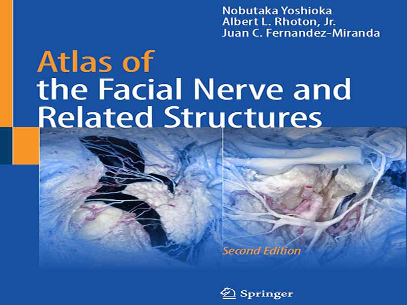
- عنوان کتاب: Atlas of the Facial Nerve and Related Structures
- نویسنده: Nobutaka Yoshioka, Albert Rhoton
- حوزه: پزشکی اطفال
- سال انتشار: 2025
- تعداد صفحه: 152
- زبان اصلی: انگلیسی
- نوع فایل: pdf
- حجم فایل: 8.13 مگابایت
حوزه جراحی احیا صورت، یکی از عمیقترین نقاط تلاقی هنر، علم و شفقت انسانی را نشان میدهد. این رشته که به دنبال بازگرداندن حرکت، بیان و هویت به افراد مبتلا به فلج صورت است، در سالهای اخیر پیشرفتهای چشمگیری داشته است. در اصل، موفقیت جراحی احیا صورت به درک عمیق از شبکه پیچیده ساختارهای عصبی-عروقی و اسکلتی-عضلانی که حرکات صورت را کنترل میکنند، بستگی دارد. در همین زمینه است که دکتر نوبوتاکا یوشیوکا، متخصص مشهور جهانی در جراحی نوروپلاستی و ترمیمی، این اطلس آناتومیک پیشگامانه را خلق کرده است. سفر دکتر یوشیوکا برای تولید این اثر به اندازه خود اطلس خارقالعاده است. دکتر یوشیوکا، دانشجوی فداکار آناتومی و جراحی، آموزشهای اولیهاش شامل مربیگری زیر نظر پروفسور افسانهای آلبرت ال. روتون جونیور، در دانشگاه فلوریدا بیش از 20 سال پیش بود و در واقع، او تنها و تنها جراح نوروپلاستی است که تاکنون چنین آموزش تخصصی را در فهرست بسیار منحصر به فرد اعضای پروفسور روتون گذرانده است. در نتیجه همکاری او با پروفسور روتون، اولین نسخه از این اطلس در سال ۲۰۱۵ منتشر شد. به طرز چشمگیری، بیش از دو دهه پس از آموزش او نزد پروفسور روتون، تعهد دکتر یوشیوکا به پیشرفت در رشته خود، او را بر آن داشت تا شش ماه را در آزمایشگاه من در دانشگاه استنفورد – مرکز آموزش و نوآوری جراحی مغز و اعصاب استنفورد – بگذراند و مطالبی را که اساس ویرایش دوم این اطلس را تشکیل میدهد، اصلاح و تکمیل کند. تلاش او برای دقت، وضوح و ارتباط جراحی در هر تشریح آناتومیکی این اطلس منعکس شده است. با این حال، این اطلس صرفاً مجموعهای از تشریحهای آناتومیکی نیست؛ بلکه یک کلاس درس در آناتومی میکروسکوپی است که به جراحی احیا صورت مربوط میشود. با توجه به افزایش اهمیت تکنیکهایی مانند پیوند عصب، انتقال عضله و بازسازی فلپ آزاد، درک کامل آناتومی صورت هرگز تا این حد حیاتی نبوده است. رویکرد دقیق دکتر یوشیوکا منبعی را ارائه میدهد که همزمان کاربردی و عمیق است و شکاف اساسی بین آناتومی نظری و تکنیک جراحی کاربردی را پر میکند. یکی از ویژگیهای بارز این اطلس، جهتگیری بالینی آن است. برخلاف مراجع آناتومیک سنتی که اغلب تشریحهای ایستا را به تصویر میکشند، این اثر بر ماهیت پویای روابط آناتومیک تأکید دارد – که برای جراحانی که احیا صورت را انجام میدهند ضروری است. تصاویر نه تنها ساختارهای مورد علاقه را برجسته میکنند، بلکه زمینه روشنی برای ارتباط آنها با روشهای جراحی خاص فراهم میکنند. مسیرهای عصبی، عروق و محلهای قرارگیری عضلات با وضوح خیرهکنندهای ارائه شدهاند و به جراحان اجازه میدهند تا راهروهای جراحی را تجسم کرده و چالشهای بالقوه را پیشبینی کنند. این اطلس فراتر از برتری بصری، گواهی بر تعهد دکتر یوشیوکا به آموزش جراحی است. این مطالب نه تنها تجربه او به عنوان یک جراح عملی، بلکه فداکاری او برای آموزش نسلهای آینده را نیز منعکس میکند. رویکرد روشمند او، تحت تأثیر آموزههای پروفسور روتون، به گونهای طراحی شده است که کنجکاوی، دقت و اعتماد به نفس را در کسانی که به دنبال تسلط بر پیچیدگیهای احیا صورت هستند، القا کند. این اثر هم یک مرجع عملی و هم یک میراث ماندگار است. با تکامل حوزه احیا صورت، تقاضا برای جراحانی با تسلط عمیق بر آناتومی زیربنایی آن نیز افزایش خواهد یافت. اطلس دکتر یوشیوکا به جراحان، دانشجویان و مربیان فرصتی بینظیر برای تعمیق درک خود از این حوزه حیاتی ارائه میدهد. از مسیرهای اعصاب جمجمهای گرفته تا پیچیدگیهای تکنیکهای انتقال عضله، این اطلس بدون شک به عنوان سنگ محکی برای برنامهریزی جراحی، آموزش و نوآوری عمل خواهد کرد.
The field of facial reanimation surgery represents one of the most profound intersections of art, science, and human compassion. This discipline, which seeks to restore movement, expression, and identity to those affected by facial paralysis, has seen remarkable advances in recent years. At its core, the success of facial reanimation surgery depends on an intimate understanding of the intricate web of neurovascular and musculoskeletal structures that govern facial movement. It is within this context that Nobutaka Yoshioka, MD, world renown expert on Neuroplastic and Reconstructive Surgery, has created this groundbreaking anatomical atlas. Dr. Yoshioka’s journey to produce this work is as extraordinary as the atlas itself. A devoted student of anatomy and surgery, Dr. Yoshioka’s formative training included mentorship under the legendary Professor Albert L. Rhoton, Jr., MD, at the University of Florida over 20 years ago, and he is, in fact, the one and only neuroplastic surgeon to ever undergone such specialized training within the highly exclusive list of Prof. Rhoton fellows. As a result of his work with Prof. Rhoton, the first edition of this atlas was published in 2015. Impressively, over two decades after his training with Prof. Rhoton, Dr. Yoshioka’s commitment to advancing his field led him to spend six months in my laboratory at Stanford University—the Stanford Neurosurgical Training and Innovation Center, refining and perfecting the material that forms the basis of the second edition of this atlas. His pursuit of precision, clarity, and surgical relevance is reflected in every anatomical dissection of this atlas. However, this atlas is not merely a collection of anatomical dissections; it is a masterclass in microsurgical anatomy as it relates to facial reanimation surgery. With the rising prominence of techniques like nerve grafting, muscle transfers, and free flap reconstructions, a thorough understanding of facial anatomy has never been more crucial. Dr. Yoshioka’s meticulous approach offers a resource that is at once practical and profound, bridging the essential gap between theoretical anatomy and applied surgical technique. A defining feature of this atlas is its clinical orientation. Unlike traditional anatomical references, which often depict static dissections, this work emphasizes the dynamic nature of anatomical relationships—essential for surgeons performing facial reanimation. The illustrations not only highlight structures of interest but also provide clear context for their relevance to specific surgical procedures. Nerve paths, vascular supply, and muscle insertions are presented with stunning clarity, allowing surgeons to visualize surgical corridors and anticipate potential challenges. Beyond its visual excellence, this atlas is a testament to Dr. Yoshioka’s commitment to surgical education. The material reflects not only his experience as a practicing surgeon but also his dedication to teaching future generations. His methodical approach, influenced by the teachings of Prof. Rhoton, is designed to inspire curiosity, precision, and confidence in those who seek to master the complexities of facial reanimation. This work is both a practical reference and an enduring legacy. As the field of facial reanimation continues to evolve, so too will the demand for surgeons with a profound command of the anatomy that underlies it. Dr. Yoshioka’s atlas offers surgeons, students, and educators a unique opportunity to deepen their understanding of this critical field. From the pathways of cranial nerves to the intricacies of muscle transfer techniques, this atlas will undoubtedly serve as a touchstone for surgical planning, education, and innovation.
این کتاب را میتوانید از لینک زیر بصورت رایگان دانلود کنید:


نظرات کاربران